

|
||
|
||
Vol. 10 (1): June 2007 |
||
Possible seal pox in the Monachus monachus Cyprus colony
|
 |
|
|
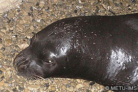
Fig. 1. Bombaci, recorded by automatic camera in a cave in Northern Cyprus. |
In July 2006, he was spotted in front of a cave on the island of Cyprus, almost 45 miles south of the cave where he was previously sighted. The large and inimitable posterior-ventral scar and identical white belly patch left no doubt as to the identity of the individual [see Seals of Northern Cyprus , TMG 9(2): 2006]. The cave was monitored with infrared monitors and more than 50 photos of Bombaci were taken automatically during the period between July 2006 and January 2007 (Fig. 1). These photographs enabled closer inspection of the scar on his body (Figs. 2 & 3).
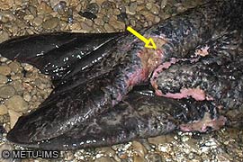
Since the bleeding lesions were considered a cause for concern, Uludag University, Faculty of Veterinary Medicine, Department of Internal Medicine, was contacted for advice and input. As the skin lesions were possibly a symptom of infection by phocine distemper virus, photos were also sent to the faculty for closer examination. Cutaneous nodular lesions on the cranial region of the rear flipper however (Fig 3) seem more like pox virus lesions, which has been identified morphologically in skin lesions of both captive and free-ranging pinnipeds and cetaceans (The Merck Veterinary Manual 2006). Seal pox is a proliferative lesion characterized by the formation of numerous 0,5 to 3 cm cutaneous nodules on the head, neck, and flippers of affected pinnipeds (Becher 2002). These nodules eventually ulcerate and are slow to heal. Cutaneous spread of this disease is mostly by head and neck rubbing, a common social behaviour of sea lions and other pinnipeds. A break in the epithelial surface is required to start an infection. Lesions can recur (Hicks 1987). The large scars around the nodules on Bombaci’s rear flipper and abdomen that developed over the healed areas may indicate that the lesions are recurring. Numerous small nodules on the head and neck are appropriate to the classical appearance of cutaneous poxvirus lesions although electron microscopy and/or PCR testing on tissue samples from lesion areas are needed to diagnose the possible agent (Tryland et al. 2005).
 |
 |
|
|
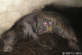
Fig. 2. Photographs recorded by automatic camera allowed closer inspection of the scar and lesions on his body. [enlarged image] |
Fig. 3. Close up image showing cutaneous nodular lesions on the cranial region of the rear flipper. |
Poxvirus is rarely fatal; however, it is regarded as having an opportunistic nature, causing outbreaks with high morbidity when the immune status of the animals are low, when food availability is scarce or when the animals are stressed (Tryland et al. 2005), as recently seen among reindeer in Finland and Norway (Büttner et al. 1995, Tryland et al. 2001).
Poxviruses from seals are zoonotic, giving cutaneous infections on fingers and hands of people handling diseased animals (Hicks & Worthy 1987, Tryland 2000), which should be noticed by persons involved in handling and care of seals.
Becher, P. 2002. Characterization of seal pox virus, a suspected member of the parapoxviruses. Arch Virol 147:1133-1140.
Büttner, M., C. Von Einem, C. McInnes and A. Oksanen. 1995. Klinik und Diagnostik einer schweren Parapocken-Epidemie beim Rentier in Finnland, Tierärztl. Prax. 23, 614–618.
Hicks, B. D. and G. A. Worthy. 1987. Sealpox in captive grey seals (Halichoerus grypus) and their handlers, J. Wildl. Dis. 23, 1–6.
Hicks, S. D. 1987. Seal pox in captive grey seals and their handlers, J. Wild. Dis., 23: 1.
Merck Veterinary Manual, The. 2006. Merck & Co., Inc, Whitehouse Station, NJ, USA.
Tryland, M. 2000. Zoonoses of arctic marine mammals, Infect. Dis. Rev. 2, 55–64.
Tryland, M., T. D. Josefsen, A. Oksanenand and A. Aschfalk. 2001. Contagious ecthyma in Norwegian semidomesticated reindeer (Rangifer tarandus tarandus), Vet. Rec. 149, 394–395.
Tryland, M., J. Klein, E.S. Nordøy and A.S. Blix. 2005. Isolation and partial characterization of a parapoxvirus isolated from a skin lesion of a Weddell seal. Virus Res. 108(1-2):83-7.
Copyright © 2007 Huseyin Cihan, Prof. Nilufer Aytug, Ali Cemal Gücü, The Monachus Guardian. All Rights Reserved |