 |
|
|
 |
||
E.J.O. Kompanje1, H. Güçlüsoy2 & P.J.H. van Bree3 Upon dissection, it was immediately apparent that the ribs were less solid than those observed in previous monk seal necropsies. The stomach contained two pieces of sea sponge, approximately 15 cm in length, two roots of Mediterranean sea grass, Posidonia oceanica, and a few of its leaves, five cephalopod beaks (currently under further study at the Aqua Products Faculty of Aegean University, Izmir, Turkey) and some parasites. Soft tissues were dissected from the skull, the long limb bones, scapulae and pelvic bones, which were subsequently cleaned by maceration (Anderson 1948). The skull and dentition appeared quite normal but some of the postcranial bones were remarkably light in weight and had small bony outgrowths (osteophytes) on the articulating surfaces (see figure 1). The low mass of the postcranial bones and marginal osteophytes in a young adult seal are typical features of osteoporosis with secondary arthrosis. Osteoporosis is a condition in which the amount and structural quality of bone tissue are reduced, commonly leading to fractures of isolated bones (osteoporotic fractures). Established factors for increased risk of osteoporosis in humans are female gender, advanced age and hyperthyroidism. Other factors that may increase the risk of osteoporosis are early natural menopause, premenopausal ostrogen deficiency, family history of osteoporosis, and a low dietary calcium intake. In humans, bone mass increases during childhood and adolescence. Thereafter, bone loss occurs in both sexes, more in females than in males. In females, spinal bone loss accelerates in relation to menopause but the pattern of postmenopausal bone loss in the hip bones is not known. The pattern of bone loss in wild mammals is not well studied. In zoo mammals, and in captive seals, bone loss – defined as osteoporosis – has been observed, especially in the axial bones, as a result of insufficient feeding (examples are found in the collection of the Zoological Museum, University of Amsterdam, and the collection of the Natural History Museum Rotterdam). In our Çesme seal, only the humeri, femora, scapula and pelvic bones could be studied, since the remainder of the skeleton was buried before the osteoporosis was diagnosed. Unfortunately, this also meant that the vertebrae of the seal were not accessible for study. Regardless, in the bones available, bone loss was particularly evident in the pelvic bones and scapula. It is well established in humans that the main cause of female osteoporosis is oetrogen defiency. Oestrogen deficiency could result from natural or premature menopause or a polycystic ovarian syndrome. In the case of our monk seal, the cause of the bone loss is difficult to determine. The animal was a young, sexually mature female, which had ovulated in recent years. Bone loss due to natural menopause is, therefore, not plausible and premature menopause appears equally unlikely. A more plausible cause in this seal is osteoporosis due to a chronic insufficient dietary calcium intake. The role of calcium intake in the etiology of osteoporosis in mammalian species has been the subject of controversy for years (e.g. Kanis 1989, Nordin & Heaney 1990). In humans, several studies have shown a relation between the consumption of low calcium dairy products and the incidence of osteoporotic fractures (e.g. Matkovic et al., 1979). Nutritionally related osteoporosis is also a well known condition in zoo animals offered low calcium "cafetaria-style diets" (Allen & Montali 1995). The mammalian skeleton contains about 99% of the calcium in the body. In order to preserve the skeleton, calcium absorption should equal the obligatory loss in faeces and urine. Sufficient calcium intake and absorption are important for the optimal development and the maintenance of the skeleton. Calcium-related problems in captive marine mammals have been mentioned by some authors (e.g. Bossart & Dierauf 1990), but not others (e.g. Worthy 1990). The IUCN Seal Specialist Group (1991) notes that "the poor condition due to lack of food as a result of overfishing" is one of the threats facing the Mediterranean monk seal. Israëls (1992) also noted that heavy fishing in areas that are important to monk seals for feeding may reduce local fish stocks to such low levels that the seals cannot obtain sufficient food. Lack of food could affect growth rates in the juvenile seals and possibly induce osteoporosis in older animals, especially in females following lactation. The poor condition of the seal we examined, and the fact that its stomach contained such unlikely food items as pieces of sponge and the roots and leaves of Posidonia oceanica, would be expected in an animal suffering from a chronic lack of food. As a substitute for fish or invertebrates – the normal monk seal diet – this animal may have started to eat whatever it found drifting at sea. Such behaviour has also been observed in a harbour porpoise Phocoena phocoena (Kastelein & Lavaleije 1992) and in a harp seal Pagophilus groenlandicus found by the first author along the Dutch coast. Of course, other causes of osteoporosis, such as diabetes, hyperthyroidism, hypoparathyroidism or renal failure, cannot be rejected on the basis of the available evidence. In monk seal research little attention is paid to the postcranial skeleton. In attempting to understand the current status of monk seals, including their limited reproductive capacity (Harwood 1987), our observations suggest that it may be important to examine the postcranial bones of other dead monk seals, and to preserve complete skeletons of these seals for detailed examination. The second author would like to thank his colleagues at the Underwater Research Society in Foça, and, in particular, Avni Gök, whose assistance made this note possible. Allen, M.E. & R.J. Montali. 1995. Nutrition and disease in zoo animals. In: R.R. Hofan et al. (herausg), Erkrankungen der Zootiere. Institut für Zoo- und Wildtierforschung: 215-232. Anderson, R.M. 1948. Methods of collecting and preserving vertebrate animals. National Museum of Canada, Bulletin no. 69: 138. Bossart, G.D. & L.A. Dierauf. 1990. Marine mammal clinical laboratory medicine. In: L.A. Dierauf (ed.), CRC Handbook of marine mammal medicine: health, disease and rehabilitation. CRC Press, Boca Raton: 1-52. Harwood, J. (ed.). 1987. Population biology of the Mediterranean monk seal in Greece. A report on research conducted by the Natural Environment Research Council's Sea Mammal Research Unit with financial assistance from the Commission of the European Communities and the International Fund for Animal Welfare. Sea Mammal Research Unit, National Environmental Research Council, Cambridge, UK: 1-72. Israëls, L.D.E. 1992. Thirty years of Mediterranean monk seal protection, a review. Nederlandse Commissie voor Internationale Natuurbescherming, Mededelingen no. 28: 1-65. IUCN Seal Specialist Group. 1991. Report on the urgent action meeting for safe-guarding the Mediterranean monk seal as a species. Texel, The Netherlands 10-11 December: 1-11. Kanis, J.A. 1989. Calcium supplementation of the diet: not justified by present evidence. British Medical Journal 298: 137-140. Kastelein, R.A. & M.S.S. Lavaleije. 1992. Foreign bodies in the stomach of a female Harbour porpoise (Phocoena phocoena) from the North sea. Aquatic Mammals 18: 40-46. Matkovic, K, K. Kostial, I. Simonovic, et al. 1979. Bone status and fracture rates in two regions of Yugoslavia. American Journal of Clinical Nutrition 32: 540-549. Nordin, B.E.C. & R.P. Heaney. 1990. Calcium supplementation of the diet: justified by present evidence. British Medical Journal 300: 1056-1060. Worthy, G.A.J. 1990. Nutritional energetics of marine mammals. In: L.A. Dierauf (ed.), CRC Handbook of marine mammal medicine: health, disease and rehabilitation. CRC Press, Boca Raton: 489-520.
MONACHUS MONACHUS FROM ÇESME, TURKEY
1 Natural History Museum, Rotterdam, P.O. Box 23452, NL-3001 KL Rotterdam
2 Underwater Research Society, P.K. 12, 35680 Foça, Izmir, Turkey
3 Zoological Museum, University of Amsterdam P.O. Box 94766, NL-1090 GT Amsterdam
On 7 March 1999, a female Mediterranean monk seal (Monachus monachus) was found dead near the town of Çesme, on the peninsula west of Izmir, Turkey. The seal was necropsied the following day by a veterinarian, Dr Avni Gök, and the second author. The seal weighed 116 kg and its 'nose to end of tail length' was 200 cm. The animal was emaciated; blubber thickness (measured at the abdomen) was 0.5 cm, and the vertebrae and hip bones were visible through the skin. Unidentified follicles were observed in both the left and the right ovaries. The large uterus suggested she had given birth before. The above information, together with the appearance of the teeth and the closure of skull sutures, all indicate the animal was a young adult.
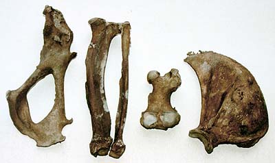
Fig. 1: Some postcranial bones were notable both for their lightness in weight and for the small bony outgrowths found on articulating surfaces.
Acknowledgements
References
![]()
![]()
![]()
![]()
![]()
Copyright © 2000 E.J.O. Kompanje, H. Güçlüsoy, P.J.H. van Bree, The Monachus Guardian. All Rights Reserved